
Imaging & Surveillance
Imaging and surveillance are important for initially identifying EHE tumors throughout the body and for assessing treatment response and/or monitoring disease changes over time. Because EHE has a high propensity for metastases and can arise in any body part or organ, whole-body imaging, typically CT, MRI, or a combination of both, is recommended at diagnosis to establish a baseline before any treatment decision is made. The imaging techniques used at baseline should be incorporated into the long-term care plan to monitor the disease and any areas of suspected spread.
Active Surveillance
Active surveillance, also called "watch and wait," is a disease management approach in which people with EHE are closely monitored through imaging, complemented by blood tests and physical exams to look for signs or symptoms of disease progression. This approach may be used to determine the need and modality of interventional treatment. Active surveillance is commonly used and recommended after diagnosis for people who are asymptomatic and not candidates for surgery, giving the care team time to assess disease progression before deciding on next steps.
This approach may feel counterintuitive to the natural instinct to take action. However, active surveillance is an intentional ongoing process of monitoring the disease.
Imaging Modalities
Assessing disease progression and treatment response are challenging in EHE. Common radiological criteria may have limited value for detecting disease progression in a timely manner, so imaging should be complemented by an assessment of disease-related signs and symptoms and patients’ quality of life. Modalities may be used independently or together.
- CT (computed tomography). CT may be used with or without a contrast dye, which makes soft tissues and blood vessels more visible. CT is grouped by body areas: head, chest, abdomen and pelvis, and spine. For liver tumors, a technique called triple-phase imaging can be used to better characterize hepatic EHE.
- MRI (magnetic resonance imaging). MRI does not use radiation and is a preferred method for assessing soft-tissue disease and for monitoring EHE in the liver and bones over time. Whole-body MRI, when available, is particularly effective at detecting bone involvement because it is sensitive to early changes.
- FDG PET/CT (fluorodeoxyglucose positron emission tomography–CT). FDG PET/CT combines a structural scan with a metabolic scan, using a small amount of injected radioactive tracer (FDG) to measure a tumor's metabolic activity. It is useful for detecting bone and limb involvement during an initial whole-body assessment.
- Bone scan. When whole-body MRI or FDG PET/CT is not available, a bone scan may be used to rule out bone involvement.
Frequency of Imaging & Scans
Imaging and scan frequency varies from person to person, depending upon individual needs and institutional care protocols.
- For disease completely removed by surgery, the Experts’ Consensus recommends imaging of the primary tumor site and a whole-body CT scan every 6 months for the first 4 to 5 years after diagnosis. Yearly scans are recommended if no disease progression is detected.
- For people on active treatment or who have suspected tumor growth, more frequent disease assessment is needed.
Measuring Tumor Change and Response to Treatment
RECIST (Response Evaluation Criteria in Solid Tumors) is a standard assessment methodology used in clinical trials and clinical practice to measure tumor response to treatment. Relying on imaging techniques such as MRI or CT, clinicians can measure tumors to detect shrinkage or growth.
However, in EHE, tumor changes may not be detectable by RECIST criteria and may not correlate with clinical benefit or overall survival. RECIST criteria focus on measurable lesions and may not account for bone metastases, ascites, or pleural effusions. Early research has demonstrated that clinical progression in EHE is associated with serosal involvement, often without meeting RECIST criteria for progressive disease. Other important factors not captured by RECIST include tumor metabolism, cellular composition, and functional characteristics.
Ongoing research aims to improve EHE assessment criteria. When assessing treatment response or determining the need for interventional therapy, specialists consider tumor response (increase or decrease in size), along with new or worsening symptoms or symptom improvement.
Related publications:
Experts’ Consensus on EHE
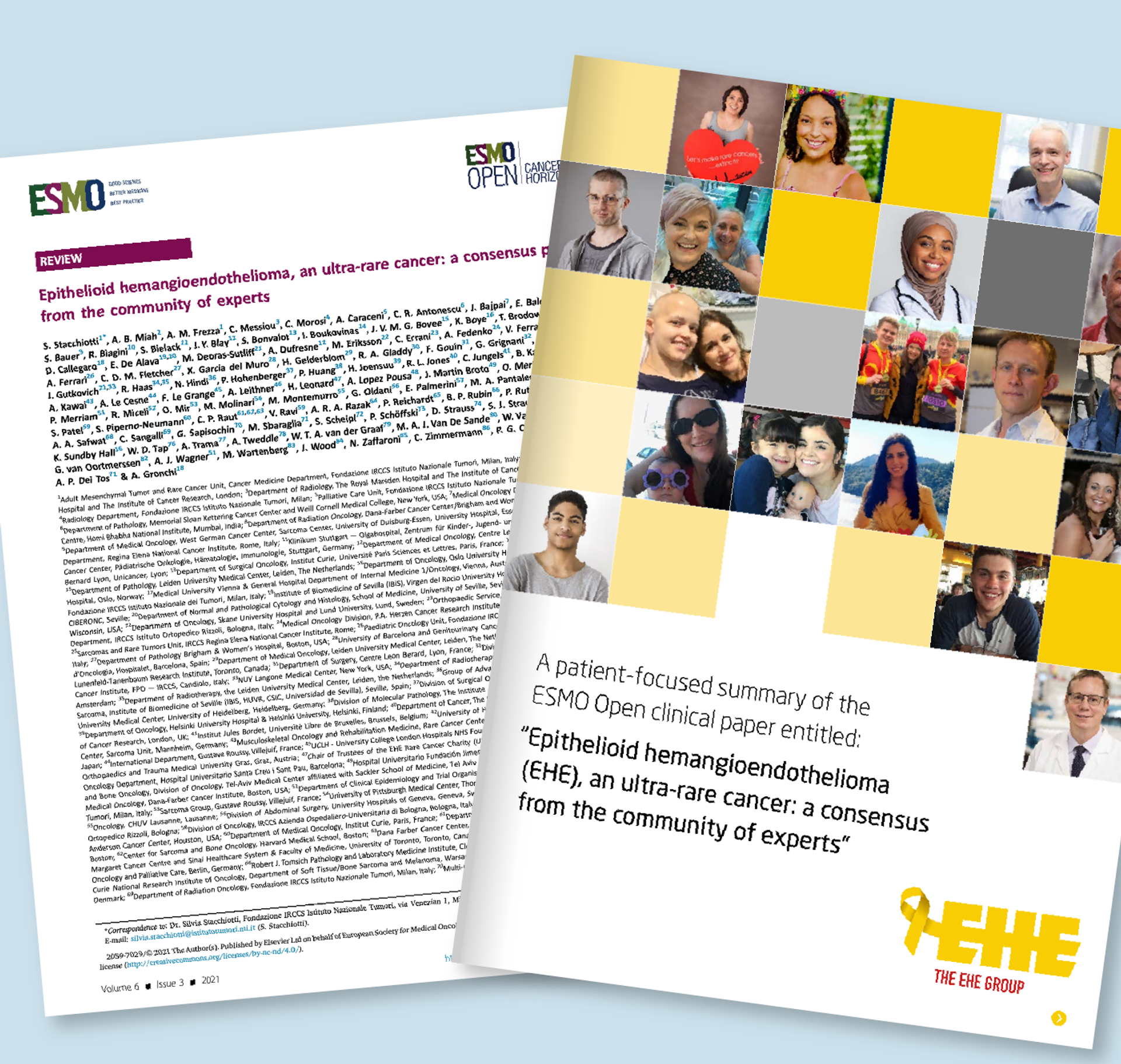
An international group of experts outlines disease biology, pathology, clinical presentation, and general principles of EHE management in the Experts’ Consensus Paper on EHE Management. Written to guide clinicians caring for people with EHE, the paper reflects current evidence and shared clinical experience across global EHE centers.
A patient-friendly version was developed to help people diagnosed with EHE understand the principles that shape care decisions. This foundational resource is a must-read for anyone affected by EHE and supports informed care planning and shared decision-making.
Continue Learning
Pain and symptom management are central to quality of life with EHE. Learn why palliative care isn’t just for the end of life.